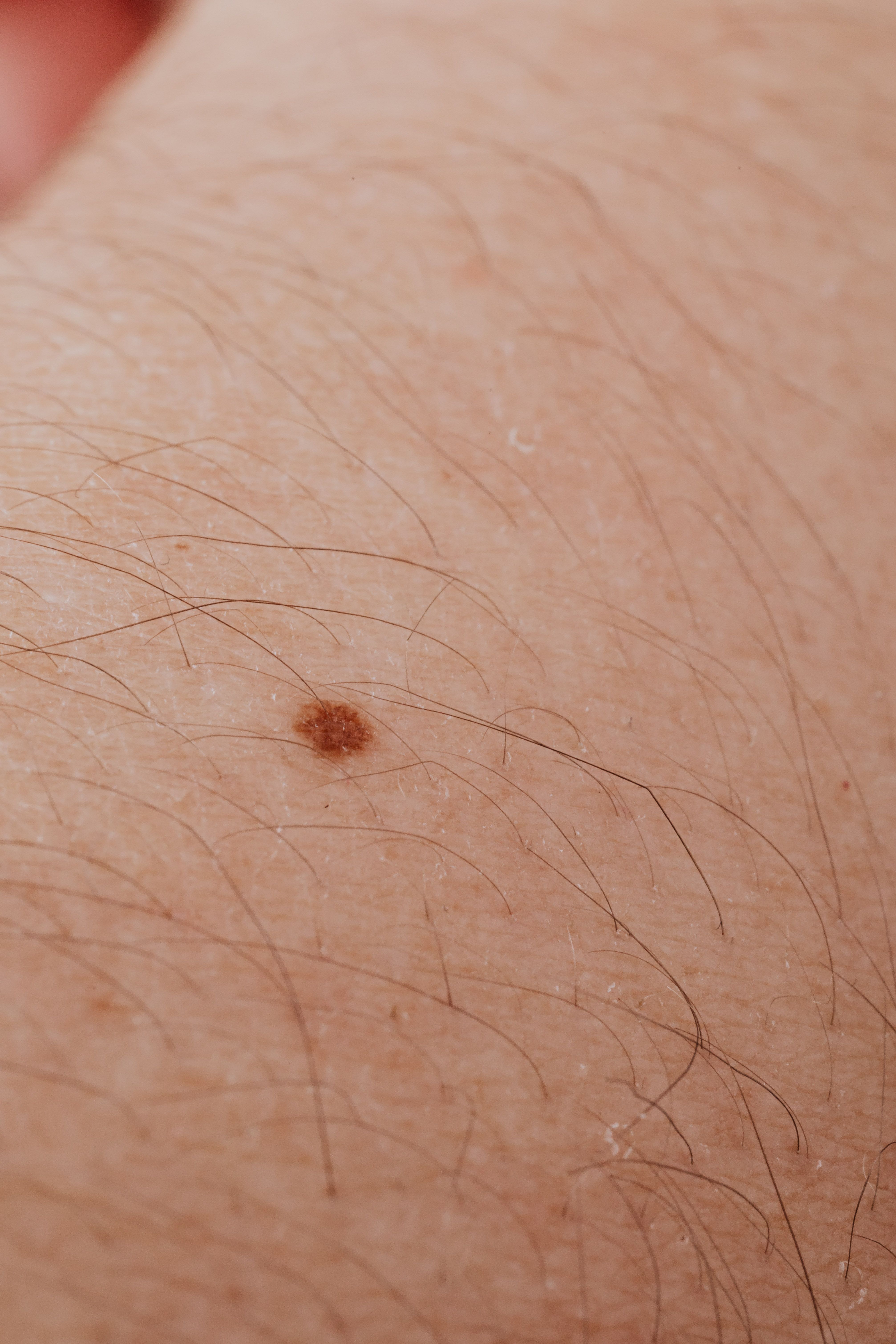
Nodular melanoma can be difficult to recognise because it may look different from the flat, irregular mole people often associate with melanoma. It may begin as a raised bump with a fairly even shape and colour.
This melanoma subtype may also grow into deeper skin layers relatively early. Recognising a new, firm, raised, or changing lesion and arranging prompt professional assessment can therefore be important.
What Should People Know About Nodular Melanoma?
Nodular melanoma is a type of melanoma that requires medical diagnosis and treatment. Information from experts can support awareness, but photographs and symptom lists cannot confirm whether a particular skin lesion is cancerous.
Important points include:
- Its border may appear fairly regular
- It may grow noticeably over time
- It may bleed or crust
- It can develop on previously normal skin
- It may not follow every ABCDE sign
- A painless lesion can still require assessment
- Dermoscopy may support clinical evaluation
- Biopsy is generally needed to confirm melanoma
- Prompt assessment matters when a lesion is changing
These features do not prove that a bump is nodular melanoma. Cysts, benign moles, inflamed follicles, and other skin conditions can also form raised lesions.
Why Is Early Nodular Melanoma Easy to Miss?
Public awareness often focuses on flat moles with irregular edges and several colours. Nodular melanoma may not match that familiar picture during its earlier development.
- It May Look Symmetrical
The ABCDE guide draws attention to asymmetry, but some nodular melanomas may initially appear round or broadly symmetrical. That appearance can make the lesion seem less concerning.
A person may therefore delay showing it to a clinician. Symmetry does not always mean harmless, particularly when a bump is new, firm, or steadily growing.
- Its Border May Appear Regular
Many people associate melanoma with jagged, notched, or poorly defined borders. A nodular lesion may have an edge that looks smoother than expected.
That feature can create false reassurance. A regular looking border cannot exclude melanoma when other changes, such as rapid growth or bleeding, are present.
- It May Have One Main Colour
Melanoma warnings often mention several colours within one lesion. Nodular melanoma may sometimes appear uniformly black, brown, red, pink, or close to the surrounding skin tone.
People can overlook lesions without obvious colour variation. A single colour is not a guarantee of safety, especially when the spot is changing.
- It Often Appears Raised
A raised bump may be mistaken for a pimple, blood blister, insect bite, cyst, or harmless mole, particularly when it first appears small.
Understanding the signs of melanoma on the palms can help people recognise concerning changes in an area that is easy to overlook.
A new or evolving lesion warrants professional assessment, especially if it grows or changes in appearance.
- It Can Develop on Normal Looking Skin
Some people expect melanoma to begin only within an existing mole. Nodular melanoma can also appear as a new lesion on skin that previously looked normal.
A new spot may not be included in someone’s usual mole checks. New growth deserves attention even when no earlier mole existed in that location.
- It May Not Cause Pain
A serious skin lesion does not need to hurt. Nodular melanoma may be painless, particularly before ulceration, bleeding, or surface irritation develops.
Waiting for discomfort can delay assessment. Visible evolution may be more important than pain when deciding whether to seek professional advice.
- It Can Grow Quickly
Nodular melanoma is associated with a more vertical growth pattern than some other melanoma forms. It can extend into deeper skin layers relatively early.
This makes delay more concerning. Noticeable growth over weeks or months should be assessed promptly, even if the lesion looks neat or evenly coloured.
Which Signs May Justify Prompt Professional Assessment?
No home checklist can diagnose nodular melanoma. A qualified clinician should assess a lesion that is new, changing, unusual, or causing concern.
Arrange an assessment for:
- A new bump that continues growing
- A firm lesion that was not present before
- A raised spot unlike surrounding moles
- A black, brown, red, or pink nodule
- A lesion that becomes larger over weeks
- Unexplained bleeding
- Repeated crusting or surface breakdown
- New itching or tenderness
- A spot that does not settle
- A lesion that changes after being photographed
- A bump mistaken for a pimple that persists
- A mole that becomes noticeably elevated
- Any skin change that worries you
- A lesion noticed by a partner or family member
The EFG guide can complement the ABCDE guide. EFG refers to a lesion that is elevated, firm, and growing, but not every nodular melanoma will display each feature clearly.
How Can You Respond to a Suspicious Raised Lesion?
The safest response is to avoid self diagnosis and arrange an appropriate clinical review. Early action allows a trained professional to examine the lesion and decide what happens next.
Record When You First Noticed It
Write down when the lesion appeared and what has changed. The timing of growth, bleeding, crusting, or discomfort can provide useful clinical context.
If possible, keep clear dated photographs. A documented timeline may show evolution that is difficult to judge from memory during one appointment.
Compare It With Other Spots
Look for a lesion that appears different from the person’s usual pattern of moles or marks. This is often described as the ugly duckling sign.
Comparison can support awareness, but it cannot provide a diagnosis. A standout lesion deserves attention even when it does not match every standard warning sign.
Avoid Picking or Treating It at Home
Do not cut, burn, squeeze, or apply unapproved removal products to a concerning lesion. These actions can cause injury, infection, and scarring.
Home treatment may also delay proper assessment. Preserving the lesion supports clinical examination and any tissue testing a qualified professional considers necessary.
Request a Professional Skin Examination
A clinician may inspect the lesion visually and use dermoscopy to examine structures not easily seen with the naked eye. They will also consider personal risk factors.
Dermoscopy can support assessment but does not replace pathology. Suspicious lesions may require biopsy or removal so tissue can be examined for a definitive diagnosis.
Share Relevant Risk Factors
Tell the clinician about previous melanoma, other skin cancers, family history, sunburn, solarium use, immune suppression, and changes in the lesion.
Risk factors do not prove that a spot is malignant. Complete information improves clinical context and can help guide examination and follow up.
Follow the Recommended Next Step
The clinician may recommend monitoring, referral, biopsy, or excision depending on the lesion. Ask when and how results will be communicated.
Do not assume silence means the result was normal. Clear follow up prevents missed action, especially when another appointment or procedure has been recommended.
Continue Checking the Rest of Your Skin
Assessment of one lesion does not remove the need to notice future changes elsewhere. Check areas that are hard to see, including the scalp and back.
Ask someone for help or use mirrors where practical. Knowing your usual skin pattern supports earlier recognition of new or evolving lesions.
What Can Support Earlier Recognition?
Awareness is most useful when it leads to timely action. People should combine skin familiarity, ultraviolet protection, and professional advice suited to their personal risk.
Practical steps include:
- Learn the normal pattern of your skin
- Notice new raised lesions
- Watch for ongoing growth
- Use the EFG guide alongside ABCDE
- Check spots that look different from others
- Photograph concerning lesions with dates
- Examine difficult areas with help
- Do not wait for pain
- Avoid removing lesions at home
- Report bleeding or repeated crusting
Skin checks cannot prevent every melanoma, but they can create opportunities for suspicious changes to be assessed earlier. Individual medical advice should guide screening frequency.
Conclusion
Nodular melanoma can be hard to recognise because it may appear symmetrical, evenly coloured, and raised rather than flat and irregular. It may also grow into deeper skin layers relatively early. A new, firm, growing, bleeding, or unusual bump should be professionally assessed. Dermoscopy may assist evaluation, but biopsy is generally required to confirm a melanoma diagnosis.


Leave a Reply