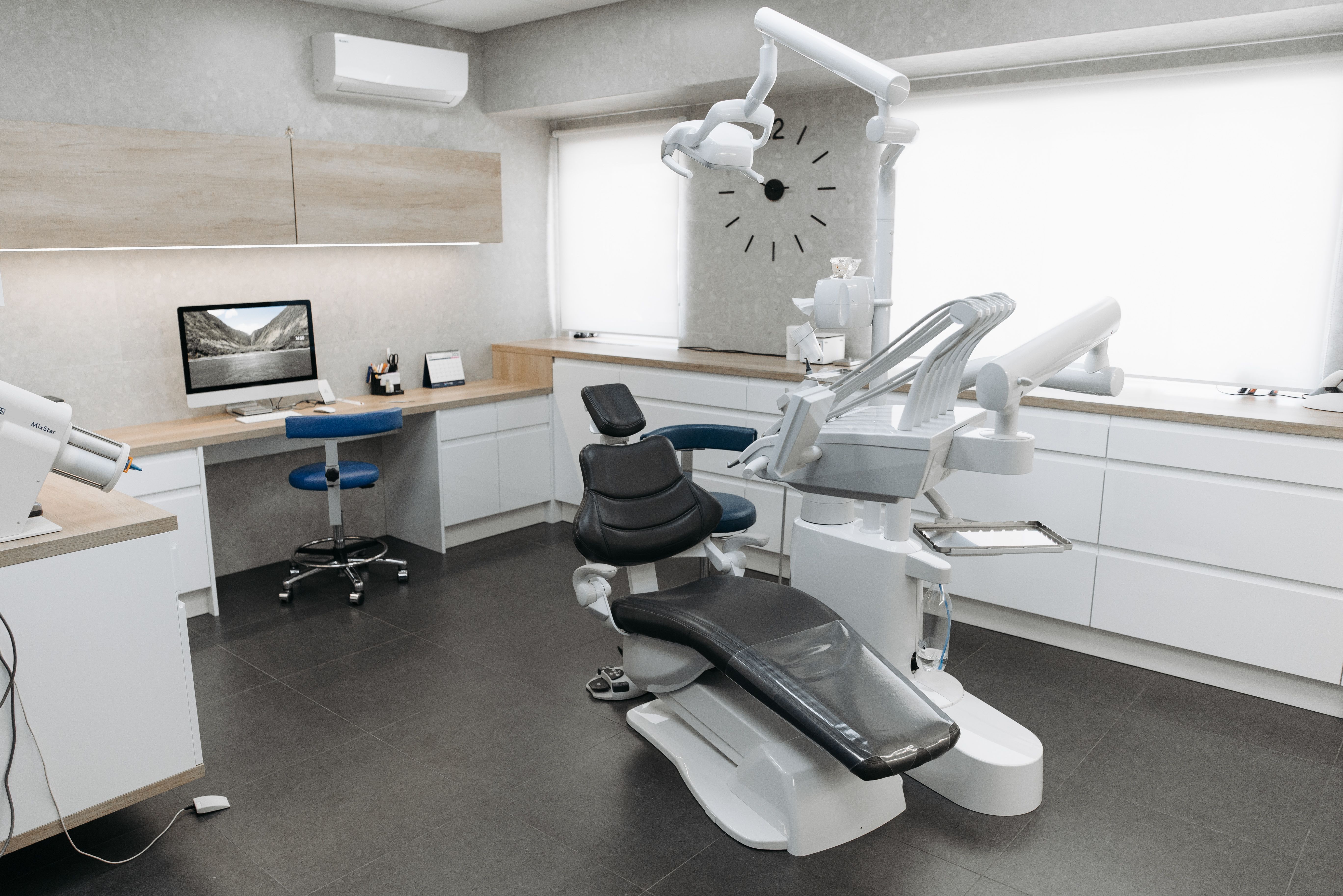
The chair still leans back. The light still shines in your eyes. But almost everything around that chair has changed in the last ten years, and most patients never get told why.
I sat in a waiting room recently and watched someone fill out a clipboard form while a tablet sat unused on the counter beside them. That gap, between the tools a practice owns and the tools it actually uses, tells you more about your visit than any brochure. Modern dentistry has quietly absorbed scanners, sensors, and software that change how your teeth get examined, how fast problems get caught, and how much of the appointment feels like guesswork. If you want to know whether your next checkup will feel different, it helps to understand what’s happening behind the mask.
The changes show up in small ways before they show up in big ones. A practice that has invested in its equipment usually tells you without being asked, and if you want to see what that looks like in practice, a dentist in winnipeg with Crestwood Dental is a useful example of how a neighborhood office frames its own technology story for patients. The point isn’t the brand name. It’s that you should be able to walk into any office and recognize what’s new, what’s old, and what it means for the two hours you’re about to spend there.
What actually changed in the chair
Three tools do most of the heavy lifting in a modern exam, and you’ve probably met at least one of them without knowing its name.
Digital X-rays replaced film in most North American practices years ago. The image appears on a screen in seconds, it can be enlarged and rotated, and the radiation dose is far lower than the old film method. If your dentist still hands you a physical film packet to hold between your fingers, you’re in a time capsule.
Intraoral scanners take the impression out of impression-taking. Instead of a tray of goop that makes you gag, a small wand sweeps across your teeth and builds a 3D model on screen. Crowns, night guards, and aligners can be designed from that model, which usually means fewer repeat visits and fewer remakes.
Cone beam imaging gives a three-dimensional view of your jaw, roots, and bone structure. It matters most for implants and complicated extractions, where a flat picture hides the parts that cause surprises.
What all three share is a shift from memory to record. Your dentist isn’t squinting at a shadow and deciding to watch it for six months. They’re comparing an image on a screen to an image from two years ago, side by side, at the same angle. That sounds minor. It isn’t.
Why catching things earlier is the real story
The technology matters because of what it does to timing. A cavity caught at the surface is a filling. A cavity caught after it reaches the nerve is a root canal and a crown, and the difference in cost and recovery is significant. Anything that helps a clinician see a problem sooner changes the trajectory of your mouth.
The same logic applies to gum health. Digital charting lets a hygienist record pocket depths consistently and compare them year over year, so a trend shows up instead of a single number. You’ve heard “your gums look a little inflamed” before. Charting turns that into “this spot went from a three to a five since last spring.” One is a vibe. The other is a decision.
Here’s the part I find most underrated: the record belongs to you as much as to the office. Practices increasingly share images and scans with patients, and that portability matters if you move, switch providers, or need a specialist. The American Dental Association has long supported patients having access to their own dental records, and digital files make that practical instead of bureaucratic.
None of this replaces a skilled set of hands. A scanner can’t tell you to floss. It just makes the conversation about flossing more specific.
The comfort side you don’t notice until it’s gone
Most patients judge a dental visit by how it felt, not by which sensor was used. Fair enough. Here’s where the technology quietly earns its keep.
Numbing delivery has improved. Computer-controlled anesthetic devices meter the flow so the pressure builds gradually, which reduces the sting of the initial pinch. It’s not painless in every case, and anyone who promises otherwise is overselling. It is noticeably less unpleasant for a lot of people.
Scheduling has changed too. Automated reminders cut the no-show rate, and a missed hygiene appointment is one of the most common reasons a small problem grows into a big one. There’s a public health angle here that rarely gets mentioned: the Centers for Disease Control and Prevention treats regular dental care as part of overall health, not a separate luxury, and anything that keeps people showing up supports that.
And then there’s the boring stuff. Better sterilization tracking, single-use barriers, and digital records of every instrument cycle. You’ll never see it, which is exactly the point.
A quick test for your next appointment
You don’t need to become an equipment expert to tell whether a practice is keeping up. Ask these four questions at your next visit and listen to how they’re answered.
- Can you show me my X-rays on the screen and explain what you’re seeing?
- Do you take digital impressions, or do we still use trays?
- Will I get a copy of my images and chart notes if I ask?
- How do you decide when to watch something versus treat it now?
The last question is the one that separates a thoughtful office from a busy one. A good clinician can explain the reasoning in plain language without a lecture. If the answer is vague, that’s information too.
| What you notice | What it usually means |
|---|---|
| Images appear on screen instantly | Digital radiography, lower dose, easy comparison over time |
| No impression trays for a crown | Intraoral scanning, fewer remakes and repeat visits |
| Hygienist types into a charting screen | Consistent pocket-depth tracking across visits |
| Reminders arrive by text automatically | Practice software reducing missed appointments |
The flip side: more data, more explaining to do
More imaging can also mean more findings, and not every finding needs a drill. A small shadow on a scan might be a cavity, or it might be a stain, a filling edge, or an artifact of the angle. The technology raises the number of things a dentist has to interpret, which puts a premium on judgment.
The U.S. Food and Drug Administration clears dental imaging devices for safety and performance, but clearance doesn’t decide what to do with the image. That call still belongs to the person holding the mirror. So when your dentist points at something on screen and recommends treatment, ask what happens if you wait three months. A confident answer, in either direction, is a good sign.
This is also where second opinions get easier. Digital files travel. If something feels rushed, you can take your images somewhere else and get a real read instead of starting from scratch.
What this means for your next visit
Technology hasn’t made dentistry automatic. It has made the information better, and better information only pays off when someone takes the time to sit with you and use it. A practice that scans your teeth but never shows you the result is just buying equipment. A practice that pulls up last year’s image next to this year’s and walks you through the difference is doing the job.
So here’s your move. At your next cleaning, ask to see your own X-rays on the screen. Not as a challenge, just out of curiosity. The way that request lands tells you almost everything about the office you’re sitting in, and it gives you a reason to pay attention to the parts of your mouth you’ve been ignoring since the last visit.


Leave a Reply